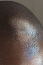
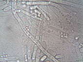
As a physician, I often explain to patients that dandruff is more than just “flakes.” Under the microscope, the picture becomes much more interesting—and helps us understand why it happens, and how to treat it effectively.

What Exactly Is Dandruff?
Dandruff is a form of scalp flaking caused by accelerated skin cell turnover. In healthy skin, cells mature and shed in about 28 days. In dandruff, this cycle speeds up to 7–14 days, leading to immature cells clumping together as visible flakes.
Microscopic Appearance of Dandruff
When scalp flakes are examined under light microscopy, several key features are often observed:
1. Clumped Keratinocytes
-
-
Dandruff flakes are made of parakeratotic cells—skin cells that have retained their nuclei due to incomplete maturation.
-
These appear as irregular, flat scales with blue-staining nuclei when viewed under special stains like hematoxylin and eosin (H&E).
-
2. Oily Coating
-
-
Sebum (skin oil) often coats the flakes, giving them a greasy appearance under the microscope. This is why dandruff sometimes feels “sticky” instead of powdery.
-
3. Fungal Elements
-
-
Malassezia spores may be visible with specific stains (e.g., PAS stain). They appear as round or oval budding yeast cells.
-
Sometimes, fungal filaments can also be seen, though Malassezia mostly exists in yeast form on the scalp.
-
4. Inflammatory Cells
-
-
In cases linked to seborrheic dermatitis, small numbers of white blood cells (lymphocytes) and damaged skin cells are visible—signs of underlying inflammation.
-
Why This Matters for Treatment
The microscopic view tells us why certain treatments work better:
-
-
Antifungal shampoos (e.g., ketoconazole, zinc pyrithione) target the Malassezia yeast seen under the microscope.
-
Keratolytic agents (salicylic acid) help break up the clumped keratinocytes.
-
Anti-inflammatory ingredients (corticosteroids, some botanical extracts) calm the immune response visible in inflammatory cases.
-
What a Doctor Looks for in Diagnosis
When a dermatologist or physician examines dandruff under a microscope:
-
-
They look at flake size, oil content, and cell structure.
-
They check for fungal elements to confirm Malassezia involvement.
-
They rule out other conditions that also cause flaking, like psoriasis (which shows thick parakeratosis with neutrophils) or eczema (which shows spongiosis).
-
Bottom Line
Dandruff may seem like a surface problem, but under the microscope, it reveals a complex interplay between skin turnover, yeast, oil, and inflammation. Understanding this microscopic picture helps guide the choice of the right shampoo or treatment—rather than relying on trial and error.
References
-
DeAngelis, Y.M., et al. Isolation and expression of a Malassezia globosa lipase gene, LIP1. J Invest Dermatol. 2007;127(9):2138-2146.
-
Pierard-Franchimont, C., et al. Dandruff: a scalp disorder triggered by Malassezia species. J Eur Acad Dermatol Venereol. 2006;20(7):760-767.
-
Schwartz, R.A., Janusz, C.A., Janniger, C.K. Seborrheic dermatitis: an overview. Am Fam Physician. 2006;74(1):125-130.